File:Fig 1a.png

Size of this preview: 603 × 600 pixels. Other resolutions: 241 × 240 pixels | 800 × 796 pixels.
Original file (800 × 796 pixels, file size: 359 KB, MIME type: image/png)
Summary
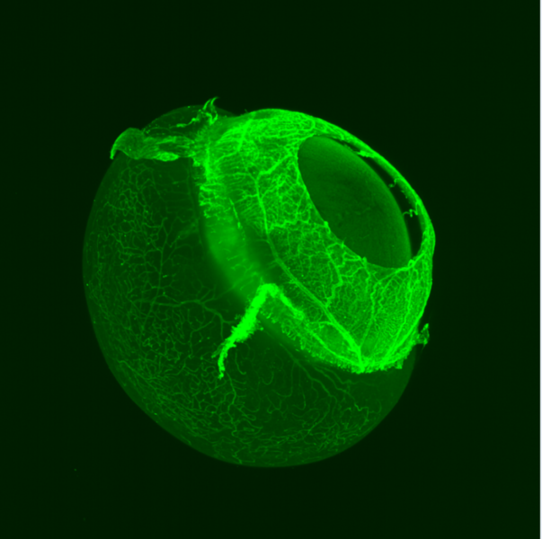
Image of mouse eyeball taken with light-sheet fluorescent microscopy, with the blood vessels shown in green.
- Prahst et al: eLife paper 2020
File history
Click on a date/time to view the file as it appeared at that time.
| Date/Time | Thumbnail | Dimensions | User | Comment | |
|---|---|---|---|---|---|
| current | 22:34, 26 May 2021 | | 800 × 796 (359 KB) | Sbprm2021 4 (talk | contribs) |
- You cannot overwrite this file.
File usage
The following page links to this file: