Difference between revisions of "Optic disk"
(→Phenotype extraction) |
(→Phenotype extraction) |
||
| Line 28: | Line 28: | ||
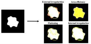
[[File:Phone.png|thumb|Figure 3. Measures of the optic disc phenotypes]] | [[File:Phone.png|thumb|Figure 3. Measures of the optic disc phenotypes]] | ||
| + | [[File:Pheno.png|thumb|Figure 3. Measure of the optic disc phenotypes]] | ||
== Phenotype extraction == | == Phenotype extraction == | ||
Once we successfully managed to isolate the optic disc from most of the pictures, our next step was to measure various of the optic disc phenotypes. We chose to focus on 4 characteristics: | Once we successfully managed to isolate the optic disc from most of the pictures, our next step was to measure various of the optic disc phenotypes. We chose to focus on 4 characteristics: | ||
Revision as of 00:10, 5 June 2023
Students: David Baumgartner, Mélissa Michel and Cécilia Torres
Assistant: Michael
Contents
Introduction
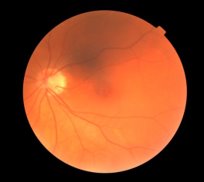
The optic disc
In the human vision, as well as in all vertebrates, there is an elliptic blind spot that is fortunately compensate by the brain so that we are unaware of its presence. This area, that has no light-sensitive cells, is called the optic disc and it is where all the signal-carrying nerve fibres are joined to become the optic nerve. The journey of visual information begins here and travels through the brain, eventually reaching the visual cortex where it will be processed and re-transmitted to other parts of the brain for interpretation.
Deep learning
Through the utilisation of artificial intelligence (AI), we can teach a computer to process data and produce accurate insights and predictions. One of the remarkable abilities of AI is its capacity to recognize pattern in complex pictures and extracts the pertinent information. In the course of this project, we initially constructed a U-Net with PyTorch. We decided to compare it with multiple models eventually choosing to switch to another model that showed better performances.
Our goal
Magnetic Resonance Imaging (MRI) procedures has a high cost and is time-consuming, we aimed to explore a less expensive and invasive alternative to extract information of the brain. Retinography seemed link a good alternative since the eyes are heavily liked to parts of the brain, also, it is comparatively a more affordable and a non-invasive procedure. This approach could allow us to approximate the brain's characteristics, thus providing a cost-effective way of obtaining preliminary information about brain structures. The primary objective of this project was to use artificial intelligence (AI) techniques to isolate the optic disc from retinography images. Then, our aim was to extract various phenotypes and characteristics of these isolated optic discs. The final objective was to find connections between the shape of the optic disc and specific regions of the brain. This work could determine if the shape of the optic disc could serve as a predictive indicator of certain brain structures.
Methods and results
Retinography segmentation
We received a hundred pictures of the eye’s back segment with their respective manually annotated images of the optic disc. These data were used to train our model, enabling it to learn to detect and isolate the accurate optic disc zone. The model was trained for 15 epochs on 100 resized 256x256 images with 5-fold cross-validation, using a binary cross entropy loss at a learning rate of 0.0001. We could go further with the project and use retinography from the UK BioBank. As we had multiple models at our disposal, our objective was to identify the one with the best performance to proceed with the project. The performances were assessed by computing the Sørensen–Dice coefficient between the labelled optic discs and the predictions of the model. After evaluation, we selected DeepLab (leftmost) as our model of choice due to its ability to deliver rapid and accurate results in comparison with the others.
However, the model still encountered challenges when processing the images. As a result, it isolated in some cases either multiple or no optic discs. To enhance the reliability of our findings, we excluded these problematic results from our research.
Phenotype extraction
Once we successfully managed to isolate the optic disc from most of the pictures, our next step was to measure various of the optic disc phenotypes. We chose to focus on 4 characteristics:
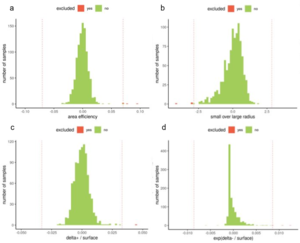
- Flattening: giving us information about the shape of the ellipse and whether it was more round or oval shaped. The flattening was calculated by fitting an ellipse on the edges of the predicted optic disc and quotiening its larger (b) and smaller radius (a). As the data follow an inverse gaussian distribution, whey were normalized by the natural logarithm minus the quotient.
- External and internal irregularities: informing us on the smoothness of the edges of the optic disc. Those were calculated by the area that were outside or inside a perfect ellipse that matched closely the actual optic disk. As we noticed that there are experimentally few cases with internal irregularities, we transformed that dataset with an exponential to better approach a normal distribution.
- Area efficiency: This trait also indicated the smoothness of the edges of the optic disc. It was calculated by the ratio of the perimeter by the surface area of the optic disc.
These four characteristics, gave us a valuable insights into the shape and smoothness of the optic disc, providing us important data for our research. Data points beyond or above 4 standard deviations of the means were excluded as outliers.
Brain traits and correlation
In order to establish correlations between the phenotypic measures of the optic disc and relevant brain regions, we specifically focused on regions that were part of the visual information pathway and had data available in the UK BioBank. We selected the optic chiasm, thalamus, lateral geniculate nucleus, and areas V1 and V2 of the visual cortex among other.
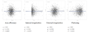
For each brain region, different types of information were accessible, including volume, mean intensity, weighted mean, area, and dispersion. For each brain trait and each optic disc phenotypes, the relation was computed as a Spearmann correlation on the residuals of a linear regression by confounding factors.Surprisingly, our results did not reveal significant correlations between the measured phenotypes of the optic disc and the selected brain regions within the visual pathway. This suggested that the shape and characteristics of the optic disc do not directly influence the properties of these brain areas.
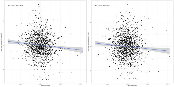
However, we discovered a correlation between the area efficiency of the optic disc and the median T2star in the substantia nigra (Spearman correlation, r² = -0.08, n = 1642, pₐⱼₛₜ = 0.03). This correlation indicated a potential relationship between the smoothness of the optic disc's edges and the median T2star value, a measure associated with tissue characteristics, in the substantia nigra. This correlation opened new investigation ideas between optic disc characteristics and the substantia nigra.
Conclusion
In conclusion, this project aimed to use artificial intelligence (AI) techniques to isolate the optic disc from retinography images and extract its phenotypic characteristics. The goal was to invesigate potential correlations between the optic disc's shape and specific brain regions, ultimately looking to predict brain structure using a less expensive and invasive procedure compared to Magnetic Resonance Imaging (MRI).
While the project did not met the expected correlations between optic disc measures and the visual pathway brain regions, it brought light on a potential relationship between the optic disc’s area efficiency and median T2star in the substantia nigra. This discovery provides new ideas for further research.
