Difference between revisions of "Cellophane"
| (19 intermediate revisions by the same user not shown) | |||
| Line 1: | Line 1: | ||
| + | [[Category:Bulletins]] | ||
| + | <newstitle> New insights in cell cycle regulation through semi-automated quantification of protein gradient </newstitle> | ||
| + | <teaser> | ||
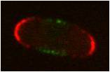
| + | In collaboration with the group of Sophie Martin from DMF (UNIL), we developed Cellophane, an ImageJ plugin that semi-automatically quantifies fluorescent protein concentration profiles along the cell cortex. This plugin enabled the quantification of hundreds of profiles of two key regulators of the fission yeast cell cycle, Pom2 and Cdr2. The data analysis, along with other experimental evidence, showed that two important functions of Pom1, deciding when and where to divide, require distinct levels of Pom1. Lower Pom1 level are sufficient for division positioning, but higher levels are required to delay mitotic entry until the proper size is reached. The paper has been published in <a href="http://www.landesbioscience.com/journals/cc/article/27411/"> Cell Cycle </a>. | ||
| + | <date>6 Dec 2013 </date> | ||
| + | </teaser> | ||
| + | |||
| + | [[Category:Homepage]] | ||
| + | |||
== Introduction== | == Introduction== | ||
| − | Cellophane | + | Cellophane is an ImageJ plugin for the semi-automated quantification of a protein profile along the cell membrane. It allows for the quantification along two channels. The plugin also has a manual mode which has a lower throughput but allows for higher precision. |
| − | [[Image: | + | [[Image:PicCellophane1.jpg| thumb | screenshot | 350px]] |
==Download== | ==Download== | ||
| − | The code is available [[Media:Cellophane.zip | here]]. This zip files contains two .java files containing the semi-automated and manual versions of Cellophane, as well as the necessary java library file. | + | The plugin is available under a GPL license and the code is available [[Media:Cellophane.zip | here]]. This zip files contains two .java files containing the semi-automated and manual versions of Cellophane, as well as the necessary java library file. |
| + | |||
| + | You can also download the short [[Media:CellophaneManual.pdf | manual]]. If you have additional questions, you can contact [[User:Micha | Micha]] | ||
== Credits== | == Credits== | ||
| − | This plugin was developed by Micha Hersch in collaboration with the lab of Sophie Martin at the University of Lausanne, in particular with Olivier Hachet. It uses the [http://www.imagescience.org/meijering/software/ | + | This plugin was developed by Micha Hersch in collaboration with the lab of Sophie Martin at the University of Lausanne, in particular with Olivier Hachet. Sascha Dalessi helped testing the software. It uses the [http://www.imagescience.org/meijering/software/ imagescience] library written by Erik Meijering. |
| + | If you use Cellophane for your published work, please acknowledge it by citing the following paper: | ||
| + | <pubmed> | ||
| + | 24316795 | ||
| + | </pubmed> | ||
Latest revision as of 12:33, 14 February 2017
Introduction
Cellophane is an ImageJ plugin for the semi-automated quantification of a protein profile along the cell membrane. It allows for the quantification along two channels. The plugin also has a manual mode which has a lower throughput but allows for higher precision.
Download
The plugin is available under a GPL license and the code is available here. This zip files contains two .java files containing the semi-automated and manual versions of Cellophane, as well as the necessary java library file.
You can also download the short manual. If you have additional questions, you can contact Micha
Credits
This plugin was developed by Micha Hersch in collaboration with the lab of Sophie Martin at the University of Lausanne, in particular with Olivier Hachet. Sascha Dalessi helped testing the software. It uses the imagescience library written by Erik Meijering. If you use Cellophane for your published work, please acknowledge it by citing the following paper:
Bhatia P, Hachet O, Hersch M, Rincon SA, Berthelot-Grosjean M, Dalessi S, Basterra L, Bergmann S, Paoletti A, Martin SG
Distinct levels in Pom1 gradients limit Cdr2 activity and localization to time and position division.
Cell Cycle: 2014, 13(4);538-52
[PubMed:24316795]
[WorldCat.org:
ISSN
ESSN
]
[DOI]
( o)
