X-rays of the structure
 and Danièle Gros (SIK|ISEA). Fabrice Ducrest © UNIL")

At the Swiss Institute for Art Research (SIK|ISEA), Danièle Gros further analysed the globes’ structure using X-ray technology. This method highlighted a number of additional details of the terrestrial globe that rounded out the results obtained using tomodensitometry at the CHUV :
- the metal shaft (axis) crossing the inside of the sphere is encased in a wooden sheath ;
- the shaft is tipped at each end by a wooden calotte ;
- the layers of the sphere are made of (or reinforced with) pieces of cloth ;
- there is an “a”-shaped piece of metal next to the southern calotte, probably serving to fasten it to the layers of paper.
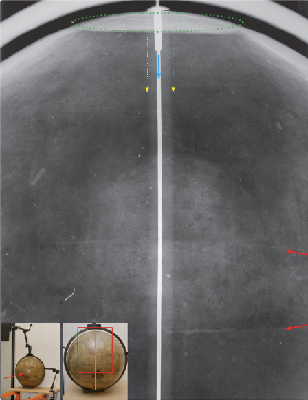
X-ray of the terrestrial globe (lower left: installation of the sphere and its position for the X-ray as seen from the machine placed one metre from the sphere. The film is just behind the sphere, pasted on the wall. The blue line indicates the position of the metal shaft and the red rectangle the area of the film). Visible on the film: the northern wooden calotte (green dots) seen almost sideways, the metal shaft (blue arrow) and wooden shaft (between the dotted yellow arrows); and the joint between the two hemispheres marked by the red arrows. © SIK|ISEA
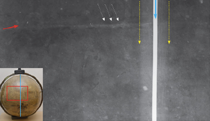
X-ray of the terrestrial globe (left-hand illustration in detail): the white dotted arrows indicate the area recognisably formed by a piece of plain-weave cloth (though this is difficult to distinguish on the image). Apparently pieces of cloth were used like the papier mâché to help make the shells. © SIK|ISEA
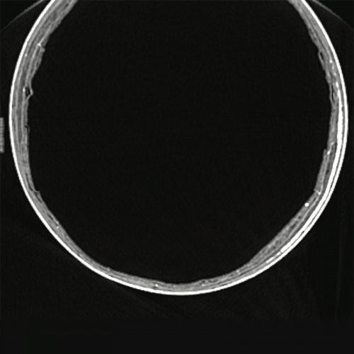
X-ray of the terrestrial globe. Cross-section of the shell, slightly towards the outer edge. The sphere is made of numerous layers which are difficult to differentiate and tend to shred in places. The chalk-paste outer layers are denser than the inner ones, which are probably made of cardboard. © SIK|ISEA
Find out more
- Les globes de Mercator de l’Université de Lausanne. Observations matérielles. Constat d’état. Rapport de conservation-restauration : The Mercator globes of the University of Lausanne. Material observations. Condition report. Report on conservation-restoration, Workshop and laboratory of the Swiss Institute for Art Research SIK|ISEA, Ref. No. 141110 0002: 01/02, October 2015, Margaux Genton, Zurich.
- Inner construction of the spheres : X-rays, computer tomodensitometry (CT) scans and 3D modelling of the resulting images help to better understand how the spheres were made.
- Shells and layers : each sphere is made of two hemispheres comprising multiple thin layers of alternating different materials. Based on binocular magnifier observations, X-rays and CT scans, the layers are made of cellulosic fibres.